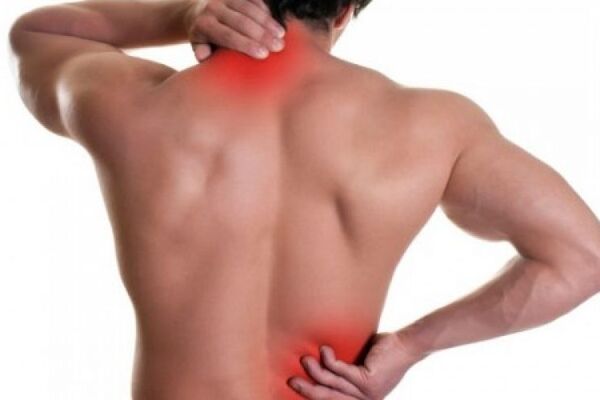
Discus hernia involves degenerative changes in the disc itself, as well as other structures of the spinal column: bone elements and ligament structure. Due to weaknesses in the prevertebral and stomach musculature, injuries, sedentary lifestyle, or lifting heavy loads, one can dislocate the intervertebral disc, shatter the anulos pulposus fibres or prolapse the nucleus pulposus. The most frequent prolapses are dorsolateral, when there is root nerve compression. Dorsomedial prolapse, when the disc pressures the spinal cord, is less frequent.
Lumbar disc herniation is most frequent between the fourth (L4) and fifth (L5) lumbar vertebrae, or the fifth lumbar (L5) and first sacral (S1) vertebrae, as the lower part of the lumbar spine carries a great portion of body weight and is under the greatest stress, and hence prone to degeneration.
With lumbar discus hernia, certain movements (like bending the torso with outstretched legs, or lifting heavy loads) can cause severe and sudden pain in the lumbar region of the spinal column that can expand down one or both legs. It can also include reduced sensitivity (numbness) in the areas affected by the root nerve compression, reduced mobility of the feet, sphincter issues (urinary and bowel disorders, and potency issues with men). Usually, there is pain during the night, and movement is severely impeded and painful. Long sitting, standing, sneezing or coughing exacerbate the pain, while lying still can help in the majority of cases.
Cervical disc herniation is a consequence of great mobility in the cervical spine, and occurs mostly between C6-C7 and C5-C6, as these vertebrae carry the greatest stress, and have a tendency to degenerate faster.
A degenerated disc is prone to shifting from its intervertebral position, which leads to neck pain if the dislodged disc pressures the posterior longitudinal ligament of the neck region of the spinal column. In the case of major disc herniation and root nerve pressuring, there is pain and weakness of the arm, followed by numbness. Extreme herniation can lead to pressures on the spinal cord, which can also manifest through leg cramps and leg fatigue, as well as urinary dysfunction. There is often a feeling of instability and disorientation, particularly in some neck movements.
In order to formulate a precise diagnosis of the condition, it is necessary to perform a radiography of the neck region of the spine (native, cervical myelography, CT scan, and magnetic resonance imaging (MRI) as the preferred method). After that, a specialist of physical therapy can formulate individual treatment plans after he performs a medical examination.
In the beginning, treatment must be non-surgical. The disc itself must be protected until its herniated portion dries out (fibrates). That is the only way to reduce its mass and release the nerves. Medication can be employed to reduce pain and inflammation, and reduce muscle spasms. They can be ingested orally, and also applied to the afflicted spot in the form of an ointment or gel. Girdles are recommended for the lumbar spine region, while the neck ought to be immobilised with a brace.
Of the available modalities of physical therapy, electric procedures, such as interference current and TENS are used in the acute stage of the affliction. Kinetic taping can reduce oversensitivity and pain in the skin or muscles, and decrease irritation of touch and pain receptors. During the subacute and chronic stages, muscular electrostimulation (classic electrostimulation, TENS, FES), ultrasound, High Intensity Laser Treatment (HILT) and massage therapy can all be applied.
Traction or spinal decompression is the only non-surgical treatment effective in severe cases of herniation, and works by relieving the affected areas of pain. It enables the majority of patients to resume their daily activities, and also to return to work, as it is quick and effective. Traction is placed at the lumbar or neck spinal region, while lying on the back, stomach or side, depending on which position the patient finds most comfortable.
Continuous horizontal positions should be avoided, except in special cases. Hence, it is very important to begin kinetic therapy as soon as the symptoms decline. It involves an individual approach and a medium-intensity programme with exercises designed to strengthen and increase the scope of movement of the paravertebral and stomach musculature in the case of lumbar disc herniation, or the neck muscles and shoulder belt in the case of cervical disc herniation. After the muscles regain their strength, the girdles will no longer be necessary.


